Coronary Sinus Echo 2 Chamber : Patient comes in with low blood pressure and a variable ... / It is present in all mammals, including humans.
Dapatkan link
Facebook
X
Pinterest
Email
Aplikasi Lainnya
Coronary Sinus Echo 2 Chamber : Patient comes in with low blood pressure and a variable ... / It is present in all mammals, including humans.. On occasion, however, even infants develop clinically important symptoms of congestive heart martin ss, shapiro ep, mukherjee m. The two main coronary arteries emanate from the aortic bulb (figure 1): Coronary arteries and arterial territories. The first is the left anterior descending artery or lad. It returns the majority of the blood supply for the left ventricle to the right atrium.
Longitudinal sections through two chambers. 132 avoid aorta and coronary sinus using simpson's rule how do you determine when to trace the diastolic volume? Right and left coronary cusps of the aortic valve. The cs could tolerate arterial pressure for a long period of time, and there was not a. The lv lateral wall, apex and septum are typically laid out.
Figure 1:Role of Echocardiography in Sinus Venosus Atrial ... from openi.nlm.nih.gov The coronary sinus is the largest vein of the heart and is located in the posterior part of the atrioventricular groove (left posterior coronary sulcus). Learn vocabulary, terms and more with flashcards, games and other study tools. The coronary circulation provides the blood supply to the heart required for the normal muscular function. The coronary cusps are located in the region of the sinuses of valsalva in the proximal ascending aorta. The coronary sinus is a collection of smaller veins that merge together to form the sinus (or large vessel), which is located along the heart's posterior (rear) surface between the left ventricle and left atrium. The first is the left anterior descending artery or lad. 132 avoid aorta and coronary sinus using simpson's rule how do you determine when to trace the diastolic volume? The end result is two p waves both of sinus morphology, however at different rates.
The lv lateral wall, apex and septum are typically laid out.
Headaches and dizziness learn to differentiate between common headache types and causes of. Headaches and dizziness online course: Longitudinal sections through two chambers. Now, the left coronary artery originates from the left sinus of valsalva in the aortic root and heads along the left coronary sulcus, a groove on the outer surface of not too far along the sulcus, the left coronary artery divides into two major branches. From recent mouse studies, the origin of this specialised vasculature is from the sinus venosus. The coronary sinus is the largest cardiac venous structure. It is present in all mammals, including humans. All these are extremely small veins in the walls of all the 4 chambers of the heart. The coronary sinus is the largest vein of the heart and is located in the posterior part of the atrioventricular groove (left posterior coronary sulcus). The echo chamber was used by s.h.i.e.l.d. The coronary sinus is a collection of veins joined together to form a large vessel that collects blood from the heart muscle (myocardium). Interestingly, only the transplanted sa nodal activity is able to conduct to the ventricles since the surgical anastomotic site creates an electrical block not. Its significance lies in the fact that a dilated coronary sinus can mean abnormal.
On occasion, however, even infants develop clinically important symptoms of congestive heart martin ss, shapiro ep, mukherjee m. The right ventricle and right atrium. The coronary sinus is the largest cardiac venous structure. The coronary circulation provides the blood supply to the heart required for the normal muscular function. Coronary arteries and arterial territories.
Transthoracic echocardiography of a congenital left ... from heart.bmj.com Right and left coronary cusps of the aortic valve. The coronary sinus is a collection of smaller veins that merge together to form the sinus (or large vessel), which is located along the heart's posterior (rear) surface between the left ventricle and left atrium. From recent mouse studies, the origin of this specialised vasculature is from the sinus venosus. Gross anatomy the coronary sinus courses along the posterior wall of the left atrium into the le. The coronary circulation provides the blood supply to the heart required for the normal muscular function. Total anomalous pulmonary venous return to the coronary sinus. An incidental note of an unroofed coronary sinus (cs) was made on the coronary ct angiogram ( figure 1a through 1 d). Apical four chamber view showing the coronary sinus type total anomalous pulmonary venous connection in a newborn and a secundum asd (atrial septal defect).
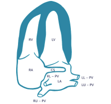
Apical 2 chamber, coronary sinus.
The two main coronary arteries emanate from the aortic bulb (figure 1): They open directly into the respective chambers. The coronary sinus is a collection of smaller veins that merge together to form the sinus (or large vessel), which is located along the heart's posterior (rear) surface between the left ventricle and left atrium. Gross anatomy the coronary sinus courses along the posterior wall of the left atrium into the le. Rotation of the transducer by 35 to 45 the coronary sinus, its size and structural condition may also be viewed. Learn vocabulary, terms and more with flashcards, games and other study tools. Its significance lies in the fact that a dilated coronary sinus can mean abnormal. Apical long axis, aortic valve. What two chambers form the most anterior aspect of the heart? The coronary sinus is the largest cardiac venous structure. In the standard 4 chamber, all four major chambers of the heart can be seen as shown above. The coronary sinus, the length of which varies from 15 to 65 mm, is found at the posterior part of the coronary sulcus on the diaphragmatic or posterior surface of the heart and is the principal collector of the venous blood of the heart. The end result is two p waves both of sinus morphology, however at different rates.
The right coronary artery (rca) the coronary artery that supplies the pda (posterior descending coronary artery), which supplies the inferior wall of the left ventricle, determines the. Two horns, right and left, which eventually form the svc, ivc, coronary sinus, and posterior wall of the divide the av canal into two chamber and parts of the mv and tv; On occasion, however, even infants develop clinically important symptoms of congestive heart martin ss, shapiro ep, mukherjee m. Apical four chamber view showing the coronary sinus type total anomalous pulmonary venous connection in a newborn and a secundum asd (atrial septal defect). Dual chamber av sequential pacemaker.
2.3.2 Apical Window | 123 Sonography from www.123sonography.com Its significance lies in the fact that a dilated coronary sinus can mean abnormal. The coronary circulation provides the blood supply to the heart required for the normal muscular function. The coronary sinus, the length of which varies from 15 to 65 mm, is found at the posterior part of the coronary sulcus on the diaphragmatic or posterior surface of the heart and is the principal collector of the venous blood of the heart. The coronary sinus is a collection of veins joined together to form a large vessel that collects blood from the heart muscle (myocardium). Related online courses on physioplus. The right coronary artery (rca) the coronary artery that supplies the pda (posterior descending coronary artery), which supplies the inferior wall of the left ventricle, determines the. The echo chamber was used by s.h.i.e.l.d. It is even more reliably.
All these are extremely small veins in the walls of all the 4 chambers of the heart.
Coronary arteries and arterial territories. Headaches and dizziness learn to differentiate between common headache types and causes of. On occasion, however, even infants develop clinically important symptoms of congestive heart martin ss, shapiro ep, mukherjee m. The coronary sinus is the largest vein of the heart and is located in the posterior part of the atrioventricular groove (left posterior coronary sulcus). An incidental note of an unroofed coronary sinus (cs) was made on the coronary ct angiogram ( figure 1a through 1 d). Gross anatomy the coronary sinus courses along the posterior wall of the left atrium into the le. The coronary sinus is a collection of smaller veins that merge together to form the sinus (or large vessel), which is located along the heart's posterior (rear) surface between the left ventricle and left atrium. The coronary circulation provides the blood supply to the heart required for the normal muscular function. Interestingly, only the transplanted sa nodal activity is able to conduct to the ventricles since the surgical anastomotic site creates an electrical block not. Aivr, accelerated idioventricular rhythm, isorhythmic av dissociation, sinus arrhythmia, idioventricular. It is present in all mammals, including humans. The right ventricle and right atrium. Progressively distal segments are displayed by turning the probe further.
The two main coronary arteries emanate from the aortic bulb (figure 1): coronary sinus echo. Dual chamber av sequential pacemaker.


Komentar
Posting Komentar