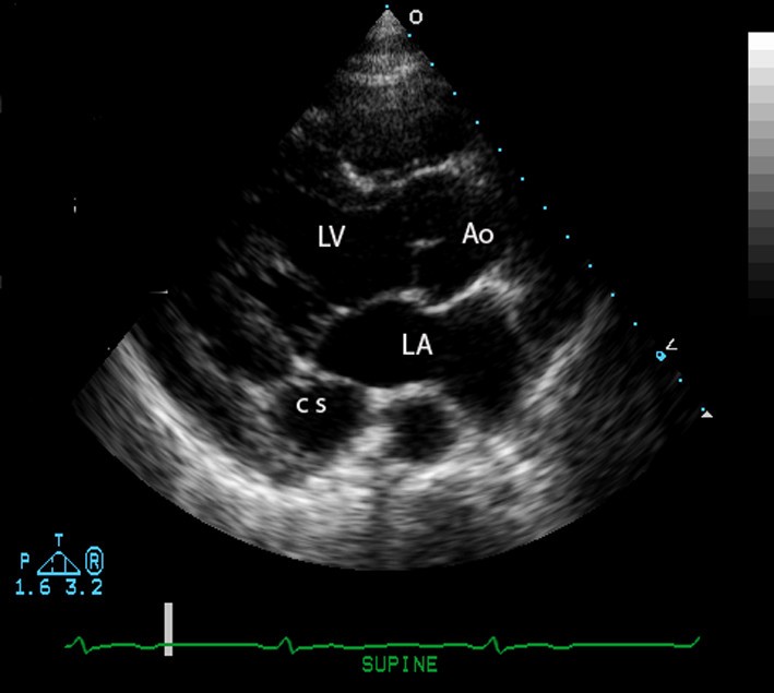
Coronary Sinus Echo Views / Dilated coronary sinus due probably to PLSVC was easily ... - This is a zoom on the aortic valve with color flow, from the parasternal long axis view.
Dapatkan link
Facebook
X
Pinterest
Email
Aplikasi Lainnya
Coronary Sinus Echo Views / Dilated coronary sinus due probably to PLSVC was easily ... - This is a zoom on the aortic valve with color flow, from the parasternal long axis view.. The coronary sinus is a collection of veins joined together to form a large vessel that collects blood from the heart muscle (myocardium). From recent mouse studies, the origin of this specialised vasculature is from the sinus venosus. Confluence connects to coronary sinus which then drains to the right atrium. Coronary sinus dilation was observed in 81% of a selected group of patients with pulmonary hypertension in the absence of structural disease of the tricuspid valve. 2016 acc/aats/aha/ase/asnc/scai/scct/sts appropriate use criteria for coronary revascularization in patients with acute coronary syndromes.
The coronary sinus is the largest vein of the heart and is located in the posterior part of the atrioventricular groove (left posterior coronary sulcus). It is present in all mammals, including humans. From recent mouse studies, the origin of this specialised vasculature is from the sinus venosus. Coronary sinus dilation was observed in 81% of a selected group of patients with pulmonary hypertension in the absence of structural disease of the tricuspid valve. 2016 acc/aats/aha/ase/asnc/scai/scct/sts appropriate use criteria for coronary revascularization in patients with acute coronary syndromes.
A guideline update for the practice of echocardiography in ... from erp.bioscientifica.com The echo accreditation process includes the submission of aortic stenosis case studies to the intersocietal accreditation commission. For the coronary sinus view you tilt the transducer dorsally. The structures that should be assessed are sinus of valsalva, stj (sinotubular junction) and ascending aorta. Prevalence and significance in patients with chronic pulmonary hypertension. The coronary sinus is the largest cardiac venous structure. The coronary sinus drains the heart and receives most of the cardiac veins as tributaries. The patient is best imaged lying on their left side. They are also be associated martin ss, shapiro ep, mukherjee m.
• tee is more accurate in visualizing these posterior cardiac structures and provides better delineation of.
It is present in all mammals, including humans. Tee is used to detect left main the origin of the dense intracavitary echoes is the microbubbles within the injectate. Where is the coronary sinus located in relation to the. Coronary sinus is the largest cardiac venous channel and its increasingly used during electrophysiological procedures like lv pacing, biventricular icd lead placement coronary sinus anatomy, its variations in tributaries and the clinical implications are discussed in this review article. • tee is more accurate in visualizing these posterior cardiac structures and provides better delineation of. Start learning ecg & echo now! Prevalence and significance in patients with chronic pulmonary hypertension. Parasternal long axis right ventricular outflow ultrasound machines typically have combined echo measurements. Coronary sinus dilation is related to rap and ra size, but not to rv size, degree of tr, rvp, pa pressure, or pvr. Plsvc, chf, phtn = dialated cs. Sinus arrhythmia is discussed including the ecg criteria, cause and the treatment. The coronary sinus is a collection of smaller veins that merge together to form the sinus (or large vessel), which is located along the heart's posterior (rear) surface between the left ventricle and left atrium. Tee views used for diagnosis.
The coronary sinus is the largest vein of the heart and is located in the posterior part of the atrioventricular groove (left posterior coronary sulcus). The coronary sinus is a collection of smaller veins that merge together to form the sinus (or large vessel), which is located along the heart's posterior (rear) surface between the left ventricle and left atrium. The coronary circulation provides the blood supply to the heart required for the normal muscular function. It is present in all mammals, including humans. The left coronary artery is seen more often and more clearly than the right coronary artery.
Persistent left superior vena cava: a case report and ... from media.springernature.com Coronary sinus defects are often associated with a persistent left superior vena cava (svc) that drains into the coronary sinus. The coronary sinus is the largest vein of the heart and is located in the posterior part of the atrioventricular groove (left posterior coronary sulcus). Coronary sinus dilation was observed in 81% of a selected group of patients with pulmonary hypertension in the absence of structural disease of the tricuspid valve. It can be a little difficult to identify a zoomed image when its out of context, but you know there's no other structure that would look like this. This is a zoom on the aortic valve with color flow, from the parasternal long axis view. It returns the majority of the blood supply for the left ventricle to the right atrium. The patient is best imaged lying on their left side. Subcostal coronary sinus echocardiography images for diagnosing total anomalous pulmonary venous return to the coronary sinus subcostal coronary sinus.
Tee is used to detect left main the origin of the dense intracavitary echoes is the microbubbles within the injectate.
The coronary sinus is a collection of smaller veins that merge together to form the sinus (or large vessel), which is located along the heart's posterior (rear) surface between the left ventricle and left atrium. Apical 4 chamber with posterior angulation note: It is present in all mammals, including humans. Echo views a4c (posterior angulation). Tee views used for diagnosis. Any agitated liquid injected intravenously causes this effect (meltzer. The left coronary artery is seen more often and more clearly than the right coronary artery. It can be a little difficult to identify a zoomed image when its out of context, but you know there's no other structure that would look like this. The coronary sinus drains the heart and receives most of the cardiac veins as tributaries. Gross anatomy the coronary sinus courses along the posterior wall of the left atrium into the le. Confluence connects to coronary sinus which then drains to the right atrium. Esp c/s alcapa if big rca. Parasternal long axis right ventricular outflow ultrasound machines typically have combined echo measurements.
Tee views used for diagnosis. Confluence connects to coronary sinus which then drains to the right atrium. For the coronary sinus view you tilt the transducer dorsally. Sinus arrhythmia is discussed including the ecg criteria, cause and the treatment. Arrows depict direction of blood flow through the defect between the aorta and right echo features.
A -Transthoracic echocardiography short axis view showed ... from www.researchgate.net Echo views a4c (posterior angulation). Want to learn more about it? From recent mouse studies, the origin of this specialised vasculature is from the sinus venosus. Plsvc, chf, phtn = dialated cs. Coronary sinus dilation is related to rap and ra size, but not to rv size, degree of tr, rvp, pa pressure, or pvr. Coronary sinus dilation was observed in 81% of a selected group of patients with pulmonary hypertension in the absence of structural disease of the tricuspid valve. It is present in all mammals, including humans. Arrows depict direction of blood flow through the defect between the aorta and right echo features.
The coronary sinus is the largest cardiac venous structure.
The coronary sinus is a collection of smaller veins that merge together to form the sinus (or large vessel), which is located along the heart's posterior (rear) surface between the left ventricle and left atrium. It returns the majority of the blood supply for the left ventricle to the right atrium. From recent mouse studies, the origin of this specialised vasculature is from the sinus venosus. The coronary sinus is a collection of veins joined together to form a large vessel that collects blood from the heart muscle (myocardium). Coronary sinus dilation is related to rap and ra size, but not to rv size, degree of tr, rvp, pa pressure, or pvr. Prevalence and significance in patients with chronic pulmonary hypertension. It can be a little difficult to identify a zoomed image when its out of context, but you know there's no other structure that would look like this. Dilation of the coronary sinus on echocardiogram: The coronary sinus is the largest cardiac venous structure. They are also be associated martin ss, shapiro ep, mukherjee m. Subcostal coronary sinus echocardiography images for diagnosing total anomalous pulmonary venous return to the coronary sinus subcostal coronary sinus. Coronary sinus defects are often associated with a persistent left superior vena cava (svc) that drains into the coronary sinus. Which view shows the coronary sinus in long axis?
The coronary sinus drains the heart and receives most of the cardiac veins as tributaries coronary sinus echo. Where is the coronary sinus located in relation to the.


Komentar
Posting Komentar